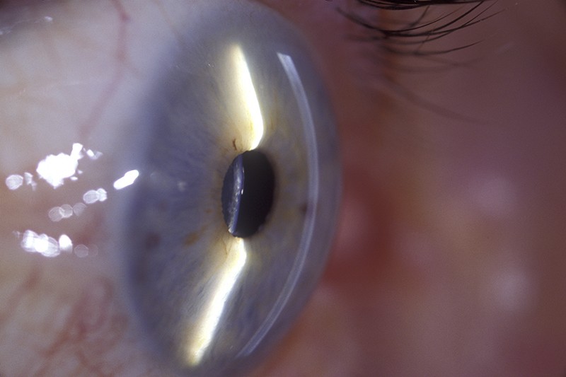
The cornea has a dampened response to prevent damage.Credit: Barraquer, Barcelona-ISM/Science Photo Library
Live-cell imaging of the eye’s transparent cornea has revealed a surprising resident — specialized immune cells that circle the tissue, ready to attack pathogens.
“We thought that the central cornea was devoid of any immune cells,” says Esen Akpek, a clinician-scientist who works on immunological diseases of the cornea at Johns Hopkins University in Baltimore, Maryland.
The study, published in Cell Reports1 on 24 May, could help researchers to better understand diseases that affect the eye and to develop therapies that target infections on the eye’s surface, says Tanima Bose, an immunologist at the pharmaceutical company Novartis in Kundl, Austria.
The cornea has a dampened response to infection, in part because aggressive immune cells could damage the clear layer of tissue and obstruct vision, says co-author Scott Mueller, an immunologist at the University of Melbourne, Australia. For this reason, the immune cells that mount a quick but crude response to an infection, such as dendritic cells and macrophages, largely reside in the outer sections of the cornea and emerge only when needed.
But in almost every tissue in the body are long-lived immune cells, known as T cells, that swiftly attack pathogens they have previously encountered — a process called ‘immune memory’. Mueller and his colleagues wondered whether such cells lived in the cornea.
Using a powerful multiphoton microscope for studying living tissue, the researchers examined the corneas of mice whose eyes had been infected with herpes simplex virus. They saw that cytotoxic T cells and T-helper cells — precursors for immune memory — had infiltrated the cornea and persisted for up to a month after the infection. Further investigations, including more intrusive microscopy techniques, revealed that the cytotoxic T cells had developed into long-lived memory cells that resided in the cornea.
The researchers then used live-cell imaging to observe the corneas of six healthy adults. They found cells similar in shape, size and speed to the patrolling T cells in mice. It was a “lightbulb moment”, says Mueller. “We were somewhat surprised and pleased to see that there is, indeed, an immune memory” in the cornea, says Mueller, who is now working on obtaining tissue from organ donors to confirm the exact type of the patrolling cells in people.
Researchers say the findings could improve the understanding of diseases such as the chronic condition dry eye, progressive corneal loss in people with autoimmune diseases, and corneal-transplant rejection.
Akpek wonders whether these long-lived immune cells are involved in shingles — a painful rash, caused by the varicella-zoster virus, that affects about one in three people in the United States during their lifetime. Some 8% of shingles cases occur in the eye, which can cause vision loss. “It makes me wonder whether there’s something wrong with the memory T cells in individuals who get recurrent shingles infections,” says Akpek.
doi: https://doi.org/10.1038/d41586-022-01578-2
Loi, J. K. et al. Cell Rep. 39, 110852 (2022).